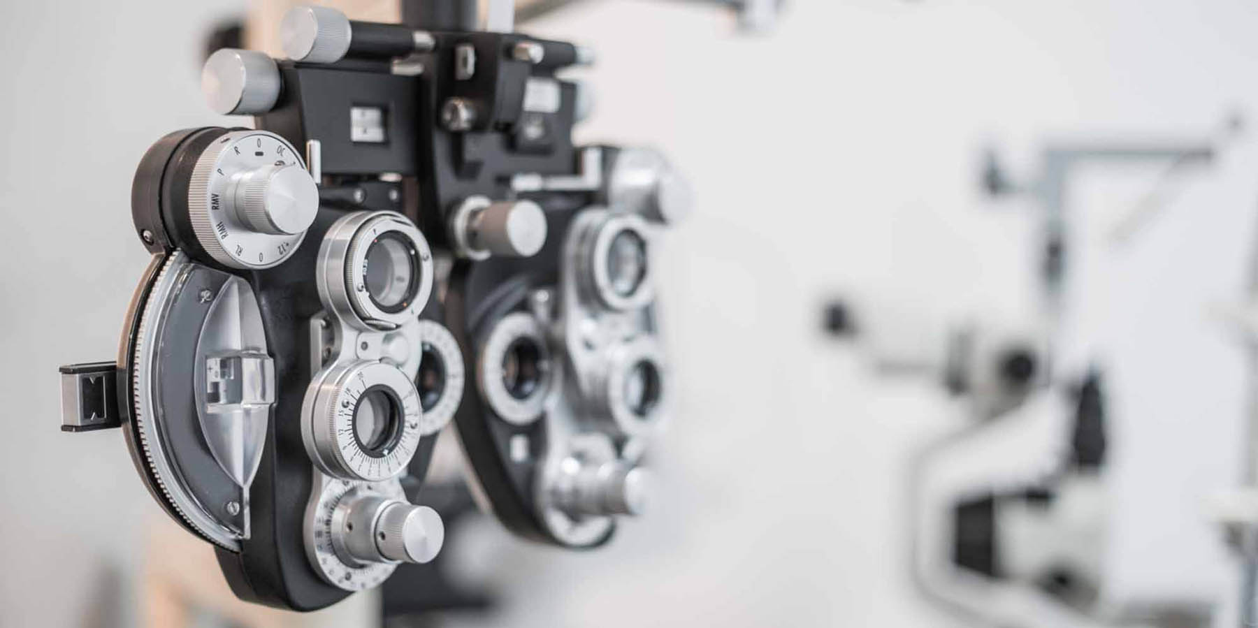
Our Technology
Accurate detection of eye disease is aided by our state-of-the-art diagnostic equipment. Since 1989, we have consistently been the first optometry practice in Hervey Bay to introduce all these new diagnostic services.
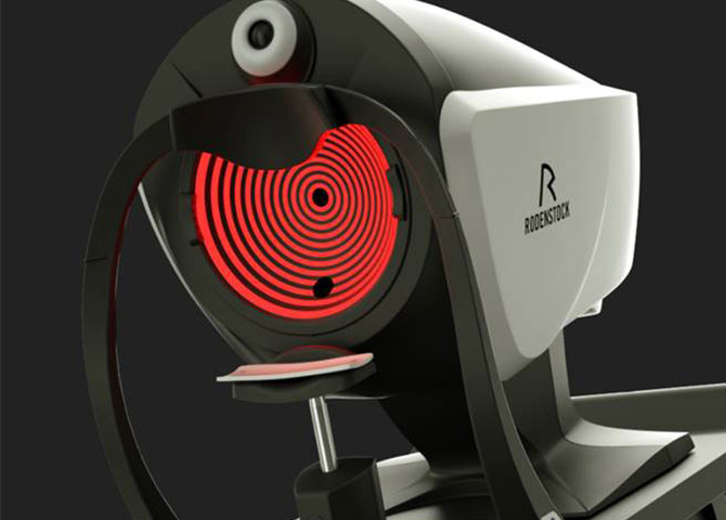
DNEye Scanner
Experience the revolutionary eyesight test with the Rodenstock DNEye® Scanner. Unique in the world with more than 7,000 measurement points transferred into your individual lens.
DNEye® optimised lenses give you the following benefits:
- High-contrast vision
- Best night vision
- Largest field of vision
- Natural visual experience
With the high-precision 3D eye measurement of the DNEye® Scanner from Rodenstock, we measure your eyes more precisely than ever before. The measured biometric values are integrated into your lenses. In doing so, you receive the most individual and perfect Rodenstock lenses and thus the sharpest vision of all time.
Available as single vision lenses and progressive lenses.
Optos Retinal Imaging
One special piece of equipment at is our Optos Optomap scanner. This equipment uses ultra-widefield (UWF™) scanning laser technology to provide a digital picture of your retina. The Optos machine can view more than 80% of the back of the eye in 3D, showing detailed views of the vessels and nerves. These images are an instant snapshot of your eye health and can be saved for future comparison to pick up any changes or anomalies.
Optos was founded by a father, Douglas Anderson, who developed the scanner for early detection of retinal detachment, after his 5 year old son Leif lost his sight in one eye when, sadly, it was diagnosed too late. His research and hard work led to the patient-friendly Optos equipment that provides the Optomap ultra wide retinal scan.
Digital Photography
Anterior: High magnification photography of the front structures of the eye aids greatly in documenting diseases and disorders such as pterygium, dry eyes, infectious conjunctivitis and keratitis, pigment changes, diseases of the cornea (front surface) and a host of other eye problems. Richard introduced this technology to Hervey Bay in 1995.
Retinal: High quality photography of the retina (light sensitive back surface of the eye) is vital in diagnosing and monitoring diabetes, macular degeneration, glaucoma, optic nerve and a raft of other posterior eye diseases. Richard introduced this technology to Hervey Bay in 1998.
Corneal Topography
Computerised corneal topography allows us to take a video image of reflections from the cornea and derive the shape and regularity of the surface.
Optical Coherence Tomography (OCT)
People often experience changes in their vision when they reach their 40s. To look out for the early signs of eye conditions, in case there’s a more serious problem, we use an Optical Coherence Tomography (OCT) device. This device is a non-invasive imaging unit that uses light waves to take cross-section pictures of your retina, the light-sensitive tissue lining the back of the eye.
Ultrasound Pachymetry
Accurate measurement of the thickness of the cornea (front surface of the eye) adds to correctly identifying glaucoma and assists with some other disorders. Richard introduced this technology to Hervey Bay in 2005.
Computerised Visual Field Testing
Accurate mapping of peripheral vision is important in the diagnosis of glaucoma, macular degeneration, pituitary disease, some strokes and a whole range of diseases of the retina (light sensitive layer of the eye). Richard introduced this technology to Hervey Bay in 1992.
Optical Biometry
Is the current standard for intraocular lens (IOL) power calculations in clinical practice and is used for measuring axial length of the eye. The process of measuring the various anatomical characteristics of the eye that are needed for IOL power calculation is called Ocular Biometry. This is particularly helpful in the management of Myopia.